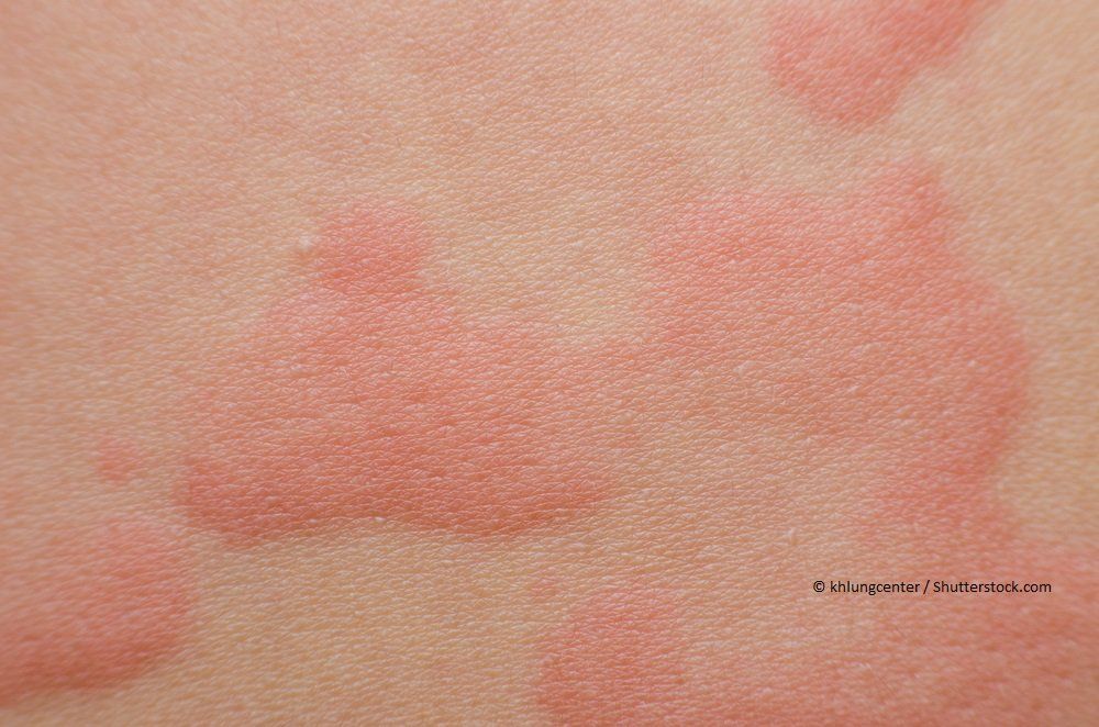

4 Treatment options for more advanced disease includes, total skin electron beam therapy, interferon therapy (IFNα and IFNγ), systemic bexarotene, extracorporeal photopheresis, biologic therapies including Alemtuzumab, a monoclonal antibody against surface antigen CD52, histone deacetylase inhibitors including vorinostat and romidepsin, as well as single and multiagent chemotherapy. 4 In cases unresponsive to the above, topical nitrogen mustard, bexarotene, local radiation, and PUVA are often employed. (See Early stage MF (Stage IA–IIA) is managed with skin directed therapies, including topical corticosteroids and phototherapy. Treatment is guided by stage and degree of body surface area involvement. Prognosis worsens in patients with extensive body surface area of involvement, those with tumor stage lesions, and as expected those with extracutaneous involvement.
Cutaneous tcel lymphoma Patch#
For those patients with limited body surface area of patch or plaque stage lesions their life expectancy is unchanged from matched controls. Prognosis depends on stage and extent of disease. In later stages of the disease, lymph nodes, hematologic, and other visceral organ involvement may occur.

The disease follows an indolent course, with progression typically occurring over many years. There is often predilection for “bathing suit” or “double covered” distribution of lesions early on in the disease course. The tumor cells are typically CD3+, CD4+, and usually CD8−. 3 Clinical presentation may be varied, with patients presenting with multiple hypopigmented to pink to hyperpigmented patches, plaques, or tumors, which may or may not ulcerate as the disease progresses. 2 Mycosis fungoides primarily affects middle age to older adults, with a recent study reporting a mean age at diagnosis of age 53.7, with a range of 8 to 91 years. Mycosis fungoides (MF) is the most common type of cutaneous T-cell lymphoma and accounts for almost 50% of all primary cutaneous lymphomas. 2 Included in this classification are Mycosis Fungoides and its variants, Sézary syndrome, CD30+ lymphoproliferative disorders, and other more rare entities including subcutaneous panniculitis-like T-cell lymphoma, extranodal NK/T-cell lymphoma nasal type, primary cutaneous peripheral T-cell lymphoma not otherwise specified, and adult T-cell leukemia/lymphoma.

Your providers might recommend evaluating your bone marrow for involvement by cutaneous lymphoma if there are any unexplained abnormal blood counts.Cutaneous T-cell lymphomas comprise approximately 75–80% of all primary cutaneous lymphomas in the Western World. SPEP screens for elevated proteins in the blood which are associated with some types of B-cell lymphomas. Hepatitis B or C testingĬarrying the Hepatitis B or C viruses can cause chronic liver inflammation, which may interfere with some treatments for cutaneous lymphoma. HTLV-1 is a virus that is common in some regions (such as Japan and the Caribbean Islands) and is associated with a subtype of CTCL. LDH is not a specific lab test, but can be elevated if there is a high rate of cells turning over in the body (such as in an aggressive cancer). Other lab tests may be ordered depending on the type and suspected stage of the cutaneous lymphoma, and include: Peripheral blood flow cytometryįlow cytometry of the blood tests for circulating lymphoma cells in the blood (also called Sézary cells), and is often done for newly diagnosed patients with CTCL. Upcoming Events for Medical Professionals.Primary Cutaneous Anaplastic Large Cell Lymphoma (PCALCL).Primary Cutaneous B-Cell Lymphoma (CBCL).

Folliculotropic Mycosis Fungoides (FMF).


 0 kommentar(er)
